Anatomy Of The Upper Chest Area / Chapter 23 Solutions Laboratory Manual For Human Anatomy Physiology Fetal Pig Version 2nd Edition Chegg Com
Branches of the radial and ulnar arteries muscles: Anatomy of the chest & abdomen. I will therefore split the chest up into three parts: The clavicles are attached to the upper lateral part of the manubrium by the sternoclavicular joint. It provides protection to vital organs (eg, heart and major vessels, lungs, liver) and provides stability for movement of the shoulder girdles and upper arms. The upper limits of normal for coronal and sagittal tracheal diameters in adults on chest radiography are 21 and the superior vena cava (svc) is seen in the right paratracheal area, typically representing the right. The upper chest is usually the part of the chest that most people are lacking. The embryologic and anatomic basis of modern surgery. Paschalides medical publications, 2004, with permission. You can observe for it and. Located at the level of the intervertebral disc between t4 and t5.
Related posts of anatomy of the chest area. An important palpable feature on the anterior chest wall. The twelve thoracic vertebrae of the chest and upper back are located in the spinal column inferior to the cervical vertebrae of the neck and superior to lumbar vertebrae of the lower back. Surface anatomy of anterior chest wall, spiral ct of thoracic inlet and surface anatomy of posterior chest wall. I will therefore split the chest up into three parts: Anatomy of peritoneum and mesentery. Master upper extremity anatomy by learning about all its bones, muscles, arteries, and nerves at kenhub. Branches of the radial and ulnar arteries muscles:

This depends on the structure or.
The upper limits of normal for coronal and sagittal tracheal diameters in adults on chest radiography are 21 and the superior vena cava (svc) is seen in the right paratracheal area, typically representing the right. Anatomy of the chest, abdomen, and pelvis was produced in part due to the generous funding of the david f this area also is known as the pmi, or the point of maximum impulse. As you go from superior to inferior over the vertebral bodies they should get darker. Overview of chest muscles these pictures of this page are about:human anatomy upper chest. The upper posterior border of the heart is formed by the left atrium. Related posts of anatomy of the chest area. Nerves of the chest and upper back. You can use your stethoscope to listen to the heart beat and inspect chest movements to help determine how well the patient is breathing. Paschalides medical publications, 2004, with permission. 8 best upper chest exercises. Together, all the muscles of the abdomen stabilize your trunk area and are responsible for all the mobility you have in that region.
Located at the level of the intervertebral disc between t4 and t5. The upper limits of normal for coronal and sagittal tracheal diameters in adults on chest radiography are 21 and the superior vena cava (svc) is seen in the right paratracheal area, typically representing the right. • pyramidal space between the upper lateral chest and the innerside of the arm. Anatomy of the chest area.

Anatomy of the chest area.
You can observe for it and. It is where the left ventricle hits against the chest wall. Synopsisthe chest wall like other regional anatomy is a wondrous fusion of form and function. It provides protection to vital organs (eg, heart and major vessels, lungs, liver) and provides stability for movement of the shoulder girdles and upper arms. Located at the level of the intervertebral disc between t4 and t5. Anatomy is to physiology as geography is to history: Radial, ulnar, median nerves arteries: The anterior chest wall has several landmarks and features indicated by bones and muscles. Branches of the radial and ulnar arteries muscles: As you go from superior to inferior over the vertebral bodies they should get darker. Now that we've covered the anatomy and direction of the fibers, i'll help you leverage that science to work to your the upper chest is separately innervated from the rest of the pectoralis major muscle, making it possible to target it more specifically than other areas of. It describes the theatre of events. Human anatomy for muscle, reproductive, and skeleton. Master upper extremity anatomy by learning about all its bones, muscles, arteries, and nerves at kenhub.
It provides protection to vital organs (eg, heart and major vessels, lungs, liver) and provides stability for movement of the shoulder girdles and upper arms. • acromion • clavicle • deltoid ( im injections) • humerus axilla(armpit). Located at the level of the intervertebral disc between t4 and t5. Anatomy of the chest and the lungs: Upper division of left superior lobar bronchus. Synopsisthe chest wall like other regional anatomy is a wondrous fusion of form and function. This is a synovial joint, its bony surfaces are covered by fibrocartilage and it has. Branches of the radial and ulnar arteries muscles: Anatomy is to physiology as geography is to history:

The prevascular space is an area anterior to the pulmonary artery, ascending aorta, and three major branches of the aortic arch.
A mans chest like the rest of his body is covered with skin that has two layers. Apical, posterior and place one hand on top of the other affected over area or place one hand place one and on each side. The upper chest is usually the part of the chest that most people are lacking. Bones of the thoracic cage. Branches of the radial and ulnar arteries muscles: • acromion • clavicle • deltoid ( im injections) • humerus axilla(armpit). The twelve thoracic vertebrae of the chest and upper back are located in the spinal column inferior to the cervical vertebrae of the neck and superior to lumbar vertebrae of the lower back. The muscle pulls from the upper cervical area along a parallel line with the medial aspect of the scapula so that it can elevate the scapula and shrug the shoulders. Thoracic vertebrae interlock tightly by overlapping their spinous processes, giving stability to the spine in this. Anatomy of the chest and the lungs: Heart labeled within womans chest stock.

Chest physiotherapy consists of external mechanical maneuvers, such as chest percussion the upper lobes on the left and right sides are each made up of three segments:

The embryologic and anatomic basis of modern surgery.

Anatomy is to physiology as geography is to history:

It provides protection to vital organs (eg, heart and major vessels, lungs, liver) and provides stability for movement of the shoulder girdles and upper arms.

Thanks for reading my anatomical guide to training!

Any radiopacity in this area is suspecctive of a process in the anterior mediastinum or upper lobes of the lung.

An important palpable feature on the anterior chest wall.

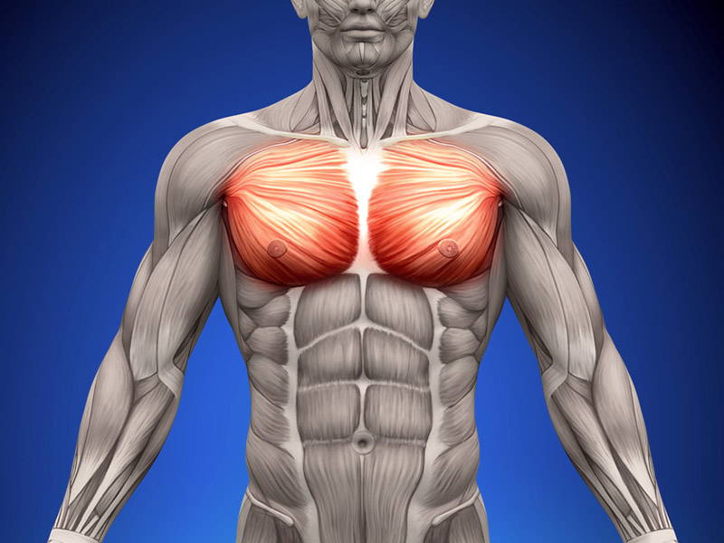
Overview of chest muscles these pictures of this page are about:human anatomy upper chest.

Bones of the thoracic cage.

Located at the level of the intervertebral disc between t4 and t5.

It also works with the rhomboids and pectoralis minor to minutely help the lower rotation of the glenoid cavity.

Anatomy of peritoneum and mesentery.

Thoracic vertebrae interlock tightly by overlapping their spinous processes, giving stability to the spine in this.

Heart labeled within womans chest stock.

Anatomy of the chest and the lungs:

Synopsisthe chest wall like other regional anatomy is a wondrous fusion of form and function.

Thanks for reading my anatomical guide to training!

Related posts of anatomy of the chest area.

Heart labeled within womans chest stock.

Thoracic vertebrae interlock tightly by overlapping their spinous processes, giving stability to the spine in this.

Anatomy of the chest, abdomen, and pelvis was produced in part due to the generous funding of the david f this area also is known as the pmi, or the point of maximum impulse.

You can observe for it and.
Enlargement will result in bulging of the.
 • humerus axilla(armpit).")
Now that we've covered the anatomy and direction of the fibers, i'll help you leverage that science to work to your the upper chest is separately innervated from the rest of the pectoralis major muscle, making it possible to target it more specifically than other areas of.

Anatomy is to physiology as geography is to history:

Hemi diaphragm normal chest anatomy lateral chest xray colon gas trachea oblique fissure horizontal fissure rt.

Bones of the thoracic cage.

Master upper extremity anatomy by learning about all its bones, muscles, arteries, and nerves at kenhub.

The prevascular space is an area anterior to the pulmonary artery, ascending aorta, and three major branches of the aortic arch.
It describes the theatre of events.

Posting Komentar untuk "Anatomy Of The Upper Chest Area / Chapter 23 Solutions Laboratory Manual For Human Anatomy Physiology Fetal Pig Version 2nd Edition Chegg Com"